
セミナー/学会・シンポジウム/市民公開講座一覧
- 検索キーワード
セミナー
第860回生医研セミナー開催のお知らせ
- 2026年11月6日(金)15:00〜16:00
- 病院馬出キャンパス 総合研究棟 104セミナー室
[対象]
内容・問合せ先内容を閉じる
内容・問合せ先
William C. Earnshaw博士 (University of Edinburgh)によるセミナーを開催します。 Earnshaw博士は、細胞分裂期染色体の構造とダイナミクスの細胞生物学者です。遺伝情報を均等に次世代へ引き継ぐ染色体安定性の分子メカニズムを解明し、近年は数理モデルと組み合わせて先端研究を展開されています。英国王立協会、米国National Academy of Sciencesのメンバーでおられます。本セミナーでは最新研究に加え、サイエンスへの情熱を聞けると思います。皆様のご来聴を心よりお待ちします。
https://www.bioreg.kyushu-u.ac.jp/mib/topics/seminar/r20261106_j.html
第860回 生医研セミナー/ The 860th MIB Seminar
日時: 2026年11月6日(金)15:00〜16:00 / 15:00〜16:00 , Friday, November 6, 2026
場所:病院馬出キャンパス 総合研究棟 104セミナー室
Seminar Room 104, 1F, Biomedical Research Station, Hospital Campus
演者:Dr. William C. Earnshaw
所属:University of Edinburgh
タイトル: Approaching Mitotic Chromosome Formation from All Directions
要旨:Attempts to understand how the DNA is packaged in mitotic chromosomes are confounded by the huge size of the DNA, the incredible chromatin density in mitotic chromosomes and the complexity of the machinery that does the DNA packaging. We study this problem by combining chemical genetics, Hi-C genomic analysis, polymer modelling, light and electron microscopy and proteomics. In our system, an entire cell population of chicken DT40 lymphocytes enters mitosis with near perfect synchrony within 2 to 3 minutes of release of a G2 phase arrest. This allows us to “kinetically section” the process and perform biochemical and structural analyses with minute-by-minute resolution. The cells can be engineered so that chromosome formation is directed by single SMC complexes: cohesin, condensin I or condensin II. Our latest models suggest that chromosomes are a disorderly helix of loops created by the SMC complexes. Condensin II drives the formation of cylindrical chromosomes but is restrained from achieving its ideal state by residual cohesive cohesin. Our electron microscopy analysis in human cells reveals that nucleosomes achieve a near millimolar concentration in mitotic chromosomes. The data from our electron microscopy and modelling are most consistent with chromosome formation involving a combination of looping by SMC complexes and chromatin phase separation. However, the chromatin concentration in chromosomes is higher than the concentration of nucleosomes in phase-separated droplets in vitro. The mechanism responsible for this compaction of the chromatin is unknown and we have recently obtained evidence inconsistent with all previous models for how the compaction is achieved. Preliminary evidence suggests that a preference of condensin I for G:C_rich DNA may drive a radial organisation where A:T-rich DNA is preferentially located towards the chromosome periphery. Despite over 140 years of study, the essential mysteries of mitotic chromosome formation remain elusive.
参考文献:
Gibcus, J.H.*, K. Samejima*, A. Goloborodko*, I. Samejima, N. Naumova, J. Nuebler, M. Kanemaki, L. Xie, J.R. Paulson, W.C. Earnshaw†, L.A. Mirny†, J. Dekker†. (2018). A pathway for mitotic chromosome formation. SCIENCE 359: eaao6135.
Cisneros-Soberanis, F.†, E. Simpson†, A.J. Beckett, N, Pucekova, S. Corless, N.Y. Kochanova, I.A. Prior, D.G. Booth*, W.C. Earnshaw*. (2024). Near Millimolar Concentration of Nucleosomes in Mitotic Chromosomes from Late Prometaphase into Anaphase, J. CELL BIOL. 223: e202403165
K. Samejima†, J.H. Gibcus†, S. Abraham‡, F. Cisneros-Soberanis‡, I. Samejima‡, A.J. Beckett, N. Pučeková, M. Alba Abad, C. Spanos, B. Medina-Pritchard, J.R. Paulson, L. Xie, A.A. Jeyaprakash, I.A. Prior, L.A. Mirny*, J. Dekker*, A. Goloborodko* & W.C. Earnshaw*. (2025). Rules of engagement for condensins and cohesins guide mitotic chromosome formation. Science 388, eadq1709.
N.Y. Kochanova†, M. Deb†, M. Borsò†, S. Webb, I. Ustun, K. Samejima, I.i Forne, I. Samejima, C. Reid, L. Xie, J.R. Paulson., A. Imhof* & W.C. Earnshaw*. (2026). A time-resolved atlas of histone modifications during mitotic entry MOL. CELL 86:1653-1672.e9.
世話人:生体防御医学研究所 がんゲノム制御学分野 斉藤典子
Noriko Saitoh, Division of Cancer Genome Biology, Medical Institute of Bioregulation
E-mail: saitoh.noriko.885@m.kyushu-u.ac.jp,
Phone: 092-642-4233
- 2026年11月7日(土)~8日(日)
- 九州大学医学部
百年講堂・同窓会会館 基礎 A 棟・総合研究棟
〒812-8582 福岡市東区馬出3丁目1番1号
[対象]ご興味のある方
内容・問合せ先内容を閉じる
内容・問合せ先
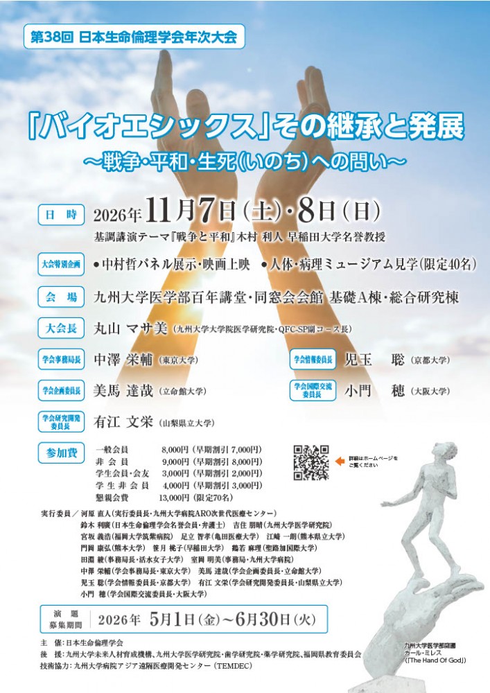
- 大会テーマ
- 「バイオエシックス」その継承と発展
~戦争・平和・生死(いのち)への問い~
- ご挨拶
-
昭和63(1988)年、日本生命倫理学会(The Japan Association For Bioethics)創立総会・記念講演会・記念パーティーが、東海大学校友会館(霞が関ビル「阿蘇の間」)において、320名の発起人と共に開催されました。
設立趣意書には、「二十世紀後半に至り科学技術の進歩は急速の度を加え、とくに、生命に関わる科学と技術の最近の発展はかつて人類が予想もし得なかった程に生命に対する知見の拡大と、また生命に対する技術的介入とを可能とするに至った。この事実は、直ちに生命現象一般に関するわれわれの基本的概念に対して重大な変革をもたらし、さらにまた、われわれの生命観・価値観に対しても根源的な反省を要請するに至った。この事実は直ちに生命現象一般に関するわれわれの基本的概念に対して重大な変革をもたらし、さらにまた、われわれの生命観、倫理観に対しても根源的な反省を要請するに至っているものと判断される。このような状況を反映して1960年代にBioethicsという概念が形成され、そして、その後、現在に至るまでの間にこの研究領域は、その対象の明確化と方法論の多様な開発とを伴って、一つの学際的な研究分野としての地位を確立するに至ったものと評価される。この時期において、日本においてもこのような世界的状況を踏まえ、多様な価値観を反映させた生命倫理に関する総合的な研究を目指す新たな学会を設立し、この分野の知見の一層の深化と拡大を計り、また、世界諸国におけるこの分野の研究との交流を促進することが緊要であると思われる。しかし、そのためには、医学、生物学を中心とする、いわゆる生命諸科学のみならず、哲学、倫理学、法学、経済学、社会学、宗教学、文化人類学など生命に係わるさまざまな学問の幅広い参加とさらにその社会的適用に係わる政策に関する学問的研究とが、同時に要請される。このような理解と判断のもとに、これら諸領域に広くまたがる学際的にして総合的な研究組織として、われわれはここに『日本生命倫理学会』の設立を提案し、志を同じくする多数の研究者の参加を希求するものである」と記された(坂本百大(2008)「『日本生命倫理学会』成立の歴史的状況―一つの記録―」『生命倫理』18(1), 4-10。このことは『バイオエシックス: その継承と発展』(丸山マサ美編著、川島書店、2018年、11頁-12頁)でも重要な事項として取り上げているところです)。
平成4(1992)年、私は、人間科学研究科修士課程より、日本生命倫理学会正会員となり、今年34年目を迎えます。
さまざまなご縁において、第38回年次大会にて、長年、教育・研究を展開した九州大学開催大会長としてのお役目を賜る事は、最高の贅沢であり、喜びでございます。
本大会では、世界のバイオエシックスを牽引された木村利人先生の基調講演『戦争と平和』をはじめ、その継承と発展における『戦争・平和・生死(いのち)』への問いを考える機会とし、また戦後81年目を迎えた今、恒久的平和を誓います。
大会企画では、1.福祉を踏まえた『動物倫理』、2.『ハンセン氏病』に触れる機会、3.『高大連携事業』と科学倫理、さらには、4.東日本大震災から15年目『原子力政策の足跡』、この企画4では、若き日の吉岡斉と『志』を同じくされた米本昌平先生をお迎えして、尊い生命観・価値観にも焦点を当てています。また、開催校オリジナル企画として、ペシャワ-ル会の協力により『中村哲医師の足跡(映画上映・パネル展示)』や人体・病理ミュ-ジアム見学も開催いたします。
本大会が、『志』を同じくする仲間との出会い・再会の機会となり、学会『設立50年』に向けた再出発の記念大会となります事を切に願い、皆さまのご参加をお待ちしております。
大会長 丸山マサ美(九州大学)
実行委員長 河原直人(九州大学)
- 開催方法
- 対面開催(一部を大会終了後に有料でオンデマンド配信)
- 会期
- 対面開催:2026年11月7日(土)・8日(日)
- 会場
- 九州大学医学部 百年講堂・同窓会会館 基礎A棟・総合研究棟
- 公式サイト
- 日本生命倫理学会の公式サイト 第38回年次大会
セミナー
第861回生医研セミナー開催のお知らせ
- 2026年11月30日(月) 10:30–11:30
10:30–11:30, Nov. 30 (Mon), 2026 - 病院キャンパス 総合研究棟 2階 ITルーム
IT Room, 2F, Biomedical Research Station, Hospital Campus
[対象]
内容・問合せ先内容を閉じる
内容・問合せ先
Harunobu Kagawa 博士によるセミナーを開催いたします。皆様のご来聴を心より歓迎いたします。
(Seminar in English)
https://www.bioreg.kyushu-u.ac.jp/mib/topics/seminar/r20261130_j.html
第861回 生医研セミナー/ The 861th MIB Seminar
日時:2026年11月30日(月)10:30–11:30 / 10:30–11:30, Nov. 30 (Mon), 2026
場所:病院キャンパス 総合研究棟 2階 ITルーム / IT Room, 2F, Biomedical Research Station, Hospital Campus
演者:Harunobu Kagawa, PhD
所属:Group Leader CR2TI-U1064 The Center for Research in Transplantation and Translational Immunology, France
タイトル:Generation of a Human Hypoblast Model to Investigate Cell Fate Specification and Self-Organization
要旨:
The hypoblast plays essential roles during early human embryogenesis by regulating epiblast development and contributing to anterior–posterior axis formation through the emergence of the anterior visceral endoderm (AVE). However, the molecular mechanisms controlling hypoblast specification and the acquisition of anterior identity remain poorly understood due to the limited accessibility of human embryos.
Here, we establish an in vitro human embryonic stem cell (hESC)-based model to investigate hypoblast differentiation and early cell fate specification. Using defined culture conditions, naïve hESCs efficiently differentiate into hypoblast-like cells expressing canonical markers including GATA4, SOX17, and PDGFRA. Time-course and molecular analyses reveal the progressive acquisition of hypoblast identity and the emergence of distinct cell populations, suggesting previously unappreciated heterogeneity during differentiation.
To investigate the signalling requirements for anterior hypoblast specification, we examined the effects of ACTIVIN and FGF/ERK signalling during differentiation. We found that sustained ACTIVIN signalling is required for the induction and maintenance of an AVE-like identity, while inhibition of MEK signalling abolishes the expression of anterior markers. These findings indicate that ACTIVIN and MEK signalling cooperate to promote the acquisition of AVE-like fate in differentiating hypoblast cells.
Together, these results establish a robust and scalable platform to investigate human hypoblast development and provide new insights into the signalling mechanisms regulating AVE-like specification. Ongoing studies using micropatterned culture systems will determine how signalling dynamics and self-organization coordinate the emergence of anterior identity during early human development.
参考文献:
- Kagawa H, Javali A, Heidari Khoei H, et al. Human blastoids model blastocyst development and implantation. Nature. 2022;601:600-605. doi: 10.1038/s41586-021-04267-8.
- Heidari Khoei H, Javali A, Kagawa H, et al. Generating human blastoids modeling blastocyst-stage embryos and implantation. Nature Protocols. 2023;18:1584-1620. doi: 10.1038/s41596-023-00802-1.
- Iyer DP, Heidari Khoei H, van der Weijden VA, Kagawa H, et al. mTOR activity paces human blastocyst stage developmental progression. Cell. 2024;187:6566-6583.e22. doi: 10.1016/j.cell.2024.08.048.
連絡先:
落合 博
生体防御医学研究所 遺伝子発現動態学分野
ochiai.hiroshi.403[@]m.kyushu-u.ac.jp
(※[]を外し半角@に変更ください。)
092-642-6882
Hiroshi Ochiai
Division of Gene Expression Dynamics, Medical Institute of Bioregulation
E-mail: ochiai.hiroshi.403[@]m.kyushu-u.ac.jp
(Please remove the square brackets and replace them with the '@' symbol.)
Tel: 092-642-6882
- 2026年12月7日(月)10:00-12:00(9:30受付開始)
- 病院キャンパス
[対象]一般の方、学生など(高校生以上)
内容・問合せ先内容を閉じる
内容・問合せ先
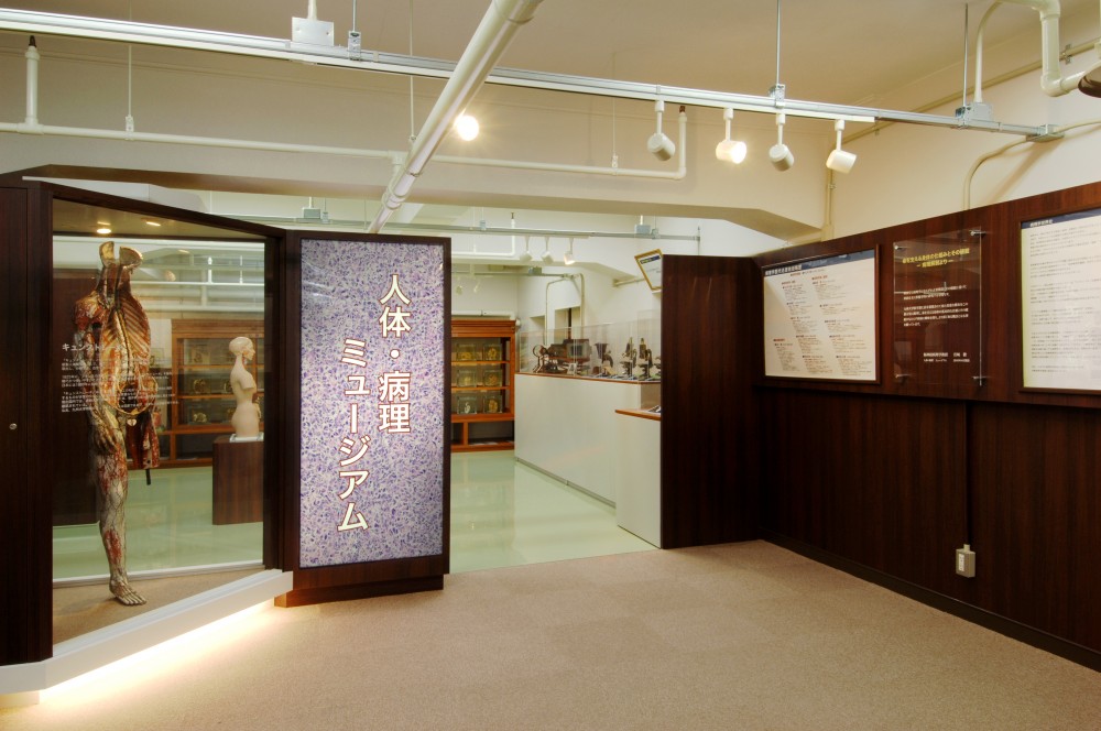
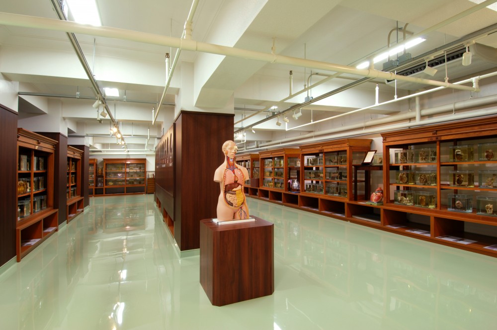
人体・病理ミュージアムの一般公開を2026年12月7日(月曜日)に実施します。
皆様のご参加をお待ちしております!
人体・病理ミュージアムとは・・・
九州大学医学部病理学教室が発足し、100年以上の時間をかけて収集した1300点の肉眼標本の中から、医学的に重要かつ希少性の高い標本をセレクトし、更に最近の興味深い標本を追加して系統的に約160点を展示しています。
また、日本国内で数体しか保存が確認されていないキュンストレーキ(紙製人体模型)や皮膚科製作のムラージュ(皮膚疾患を蝋模型にしたもの)など・・・教室の枠を超えた様々な医学史的遺産や、病理とは切り離せない顕微鏡も単眼のものから何とも形容しがたい外観の顕微鏡まで・・・骨董的価値のあるものをセレクトし展示しています。
モニターを設置した視聴覚エリアでは、病院で病理学がどのような役割を果たしているか “診断” をキーワードに紹介をしています。
その他に、九州大学病院及び病理学教室史を大型写真パネルで掲示、学内に散らばっていた史料の中から厳選した写真を中心に作成しており、100年の移り変わりを写真で見ることは、歴史をとてもリアルに感じ、まるで “タイムスリップ” したかのようです。
「百聞は一見に如かず」という諺があります。貴重な「一見」を人体・病理ミュージアムで体験しませんか?
剖検にご協力いただいた多くのご遺体のご厚情による肉眼標本の見学にあたっては、礼意を持って臨んでいただき、貴重な機会であることをご理解いただきたく、よろしくお願いいたします。
■日時
2026年12月7日(月曜日)10:00~12:00(9:30~10:00受付時間)
■場所
〒812-8582 福岡県福岡市東区馬出3丁目1-1
九州大学 病院(馬出)キャンパス 医学部基礎研究A棟(図上上の①番の建物)
九州大学医学部ホームページ https://www.med.kyushu-u.ac.jp/campus/access/
■対象
一般の方、学生など 30名
※安全及び正しい理解、冷静に見学いただくため高校生以上に限定いたします
※九州大学の教職員及び学生も対象です
■申込
事前予約制、先着順、参加無料
092-642-6073 (平日10-16時)
受付期間 10/1(木)9:00~12/7(月)9:00
申込詳細はミュージアムHPをご覧ください。
■見学内容
・自由見学(九大病院・医学部の歴史、ムラージュ、キュンストレーキ、肉眼臓器標本、人体骨標本など)
・ビデオ鑑賞(病院での”病理診断”について)
・病理スタッフによる説明ガイド及び質問対応
・詳細な展示物はミュージアムHPをご覧ください。
■詳細はミュージアムHPもしくは資料をご覧ください。
人体・病理ミュージアムHP https://byori-bohken.med.kyushu-u.ac.jp/museum.html
第13回人体・病理ミュージアム一般公開
